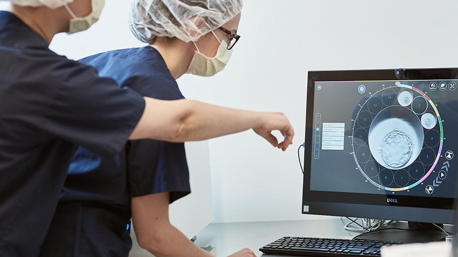
Observing embryo development
Fertilys recently acquired an incubator with an integrated time-lapse system. With this state-of-the-art technology, the embryos remain in a stable environment and there is no longer any need to remove them from their incubator to observe their continuous development.
This technology makes it possible to:
- Take pictures of the embryos every 5 to 10 minutes. View the recorded photos, when more information is desired on the development of the embryo.
- Observe important events in the embryonic development, such as fertilization and cell division.
- Know the precise time of cell division for each embryo.
- Compare the kinetic recordings of the development of several embryos to select the dominant one for transfer.
- Cultivate the embryos of a single patient per incubator chamber. There are six in total.
Even though there are no studies that show concrete evidence of an increase in pregnancy rates, the time-lapse system helps us understand considerably better the kinetics of embryo development, as it stabilizes the parameters necessary for their growth (temperature, oxygen, carbon dioxide) in the lab and reduces human error.
